Introduction:
Radiology department was established at government hospital, Salem in 1953. Initially X-ray unit was installed in this hospital and x-ray services were provided to the patients.
Later in 1989 Ultrasound service came to the radiology department, now an average of 180 to 200 cases of ultrasound studies is done daily in Radio-diagnosis department Now , we have around 10 ultrasound machines all with doppler facilities.
Doppler ultrasound with high frequency ultrasound facility started in our department In 2004, now an average of 7 to 9 scanS of doppler studies are done per day.
X-ray mammogram was started in this hospital in the year 2005. since then it is functioning effectively. An average of 4 to 5 cases of mammogram studies are done.
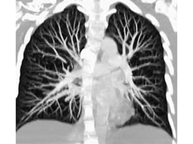
First single slice CT scan started in this hospital in 1997. Then onwards all types of CT scan taken to the both IP and OP patients, now an average 190 to 230 scans done per day. 24 hours CT scan emergency service available in this department. Now we have two 16 slice CT scanner machines and a 128 slice CT scanner machines for
the needs of the patients.
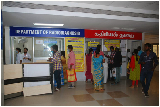
In the year 2011 the Department of Radio- diagnosis was shifted to the new super
speciality building (PMSSY building). New high quality services were established to serve the patients need.
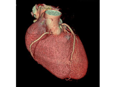
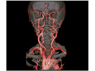
FIRST TIME in Tamilnadu 128 slice CT scan was installed in the Department of Radio diagnosis, GMKMCH Salem in the year 2011.Itprovides all types of CT angiogram studies to the patients high frequency 800mA x-ray unit with IITV was started in 2011 at Radio diagnosis department. Contrast procedure services are provided to the patients. Now an average 4 to 5 cases are done per day.
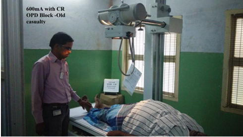
600mA x-ray unit with Computer Radiography (CR) was started at OPD block in 2015
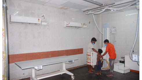
800mA X-ray unit with Digital Radiography and PACS was installed at PMSSY block in 2015
New 16 slice CT scan machine was installed in medical block during december 2019.
The installation coincided with COVID 19 outbreak. The CT machine provided
immense support in diagnosing and triaging thousands of COVID patients.
The first 1.5 Tesla MRI with higher software facilities installed in 2011 was replaced
with a new 1.5Tesla MRI machine in the year 2021 with all latest applications
to facilitate state of art imaging.
It provides all newer imaging techniques and services to the patients. On an average 25 to 30 MRI studies are done per day.
Two 3D 4D ultra sound machine with elastographyinstalled in 2021
The new 800mA IITV with DR FLUROSCOPY was installed in the year 2022 in the
medical block and is functioning effectively.
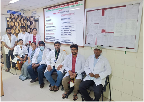
Faculty Details :
| S. No. | Designation | Name |
|---|---|---|
| 1 | PROFESSOR AND HOD | DR.P.KUMAR MD RD., |
| 2 | ASSOCIATE PROFESSORS |
DR.N.SUNDARESWARAN MDRD.,
DR.M.SANKAR MDRD., |
| 3 | ASSISTANT PROFESSORS | DR.E.ELANCHEZHIAN MDRD., DR I.GOMATHIPONSHANKAR MDRD., DNB., DR.E.VIDHYA DMRD., DNB., |
| 4 | SENIOR RESIDENTS |
DR.N.SHANTHALA MDRD., DR.A.S.KANNAN MDRD., DR.B.S.VADIVU DMRD., DR.K.THIYAGARAJAN DMRD., DR.A.DHEEPA DMRD., DR.G.JAYSIYADMRD., |
Address For Communication:
Head of the department
Department of Radio diagnosis
GMKMCH , Salem - 1
Email id: radiodiagnosissalemgh@gmail.com
Registrar: DR.M.SANKAR MDRD.,
Ph:9952425775
Other Staffs:
| S. No. | Name | Designation |
|---|---|---|
| 1 | MR.G.MUTHUTHIRUMARAN | CHIEF X-RAY TECHNICIAN |
| 2 | MR.G.THIAGARAJAN | RADIOGRAPHER |
| 3 | MR.V.KANNAN | RADIOGRAPHER |
| 4 | MRS.M.CHITRA | RADIOGRAPHER |
| 5 | MR.M.KANNAN | RADIOGRAPHER |
| 6 | MR.J.KASIVISWANATH | RADIOGRAPHER |
| 7 | MR.R.SIVA | RADIOGRAPHER |
| 8 | MRS.P.DHIVYA BHARATHI | RADIOGRAPHER |
| 9 | MISS.R.KUDIYARASU | RADIOGRAPHER |
| 10 | MRS.A.UMAMAHESHWARI | RADIOGRAPHER |
| 11 | MRS.M.SATHYA | RADIOGRAPHER |
| 12 | MRS.S.AKILA | RADIOGRAPHER |
| 13 | MR.R.VENKATESH | RADIOGRAPHER |
| 14 | MRS.M.PUSHPAVALLI | RADIOGRAPHER |
| 15 | MRS.R.AMBIGA | RADIOGRAPHER |
| 16 | MR.U.BALAJI | DARK ROOM ASSISTANT (DRA) |
| 17 | MRS.M.REVATHY | DATA ENTRY OPERATOR / STENOGRAPHER |
| 18 | MR.S.DHINAKARAN | RECORD CLERK |
Services Offered:
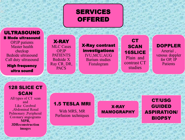
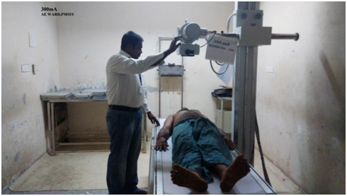
1) X ray :
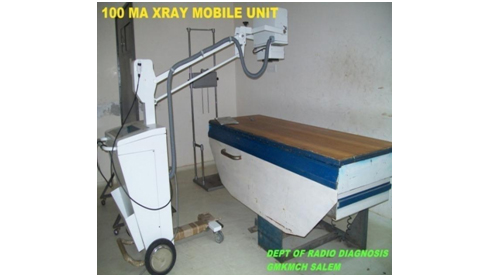
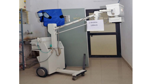
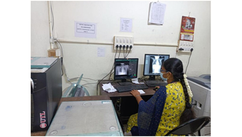
We have 4 fixed x ray units and 6 mobile x ray units. On an average 400 to 450 X rays are being taken per day.
| Machine | Strength | Fixed | Mobile |
|---|---|---|---|
| 800mA WITH IITV RECORDING SYSTEM | 800mA | 2 | - |
| 800mA DIGITAL RADIOGRAPHY - CEG - 1 | 800mA | 1 | - |
| 500mA with CR systems | 500mA | 2 | - |
| 300m with CR System | 300mA | 2 | - |
| 100mA mobile unit | 100mA | - | 6 |
2) Fluroscopy:
A well equippedfluroscopy unit - 800 mA Xray unit with image intensifier tube was established on 2011. Contrast procedures including barium studies, urographic studies are being done. An average of 5 to 8cases are done per day.
Barium studies includes : barium swallow, barium meal , barium follow through and barium enema
Urographicproceduces: IVU, MCU
LOOPOGRAM, T TUBE cholangiography
The new 800mA DR fluroscopy with IITV is installed during the year 2022. since then contrast procedures are routinely done.
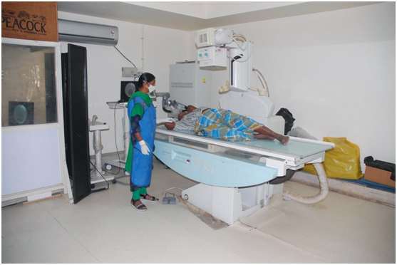
800mA with IITV at PMSSY BLOCK
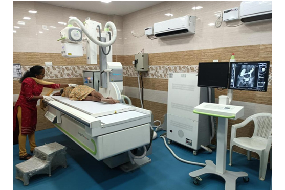
800mA DR FLUROSCOPY at medical block
3) Mammography:
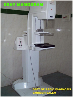
With the subsequent installation of Mammography unit . We are performing mammogram procedures on an average of 5 to 6 cases per day.
4) Conventional angiography:
Conventional angiography procedures are regulary done in our hospital. An average of 1 t o 2 cases per day are being done.
5) Ultrasound:
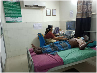
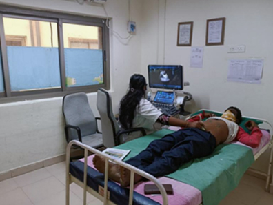
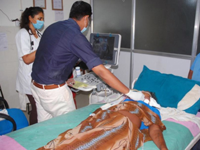
There are around 9 to 10 well equipped ultrasound DOPPLER machines with high frquency probes are available in our department . Now an average of 150 to 200 cases of ultrasound are done routinely.
We render seperate ULTRASOUND services for Out patients, Inpatients, Antenatal patients , doppler studies, superspeciality IN & OUT patients , emergency - trauma care services and USG guided biopsy - procedures.
Two advanced MINDRAY DC80 machines installed in 2021 with 4D probe and recent elastography techniques has supported the patients in diagnosing better.
ULTRASOUND
6) CT:
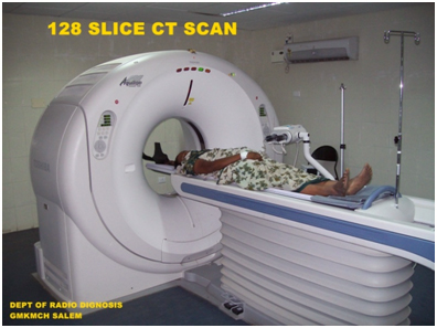
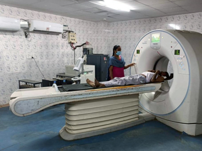
First single slice CT scan was started in 1997 followed by the commeration of 16slice CT , State of art 128 slice CT and another 16 slice CT in the subsequent 20 years.
We perform CT studies for routine as well as emergency and trauma cases. Our medical college hospital is the the prime tertiary care centre with all well establishedsuperspecialities in the northwestern Tamilnadu. We render services to the neighbouring 6 districts like Namakkal, Karur, Krishnagiri, Dharmapuri and Kallakurichi
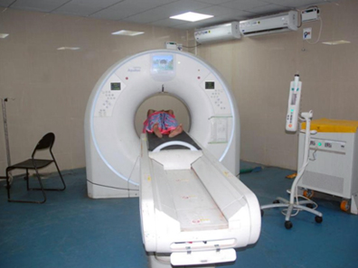
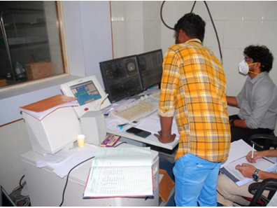
The well equipped 16slice CT scanner in the superspeciality block with the dedicated workstation functions 24 X 7days . On an average we perform 200 CT scans per day including more than 15 to 20 CT CONTRAST studies per day.
In the eve of COVID 19 pandemic ; the newly installed 16 slice CT(date of installation - 06 /12/19) scan machine in medical block is completely dedicated to scan COVID patients and we have performed more than 12,000 CT scan studies within a span of a year.
We routinely perform CT guided biopsies and interventional procedures in our department.
| S. NO | MACHINE | COMPANY | LOCATION |
|---|---|---|---|
| 1 | CT 128 SLICE | TOSHIBA Aquillon CX TSX | 114 PMSSY BLOCK RD Department |
| 2 | CT 16SLICE | Aquillon lightening 110 PMSSY | BLOCK RD Department |
| 3 | CT 16SLICE | Aquillon lightening | MEDICAL BLOCK |
7) MRI:
Our department is well equipped with a state of art 1.5tesla MRI scanner with a dedicated workstation since 2011.
The old MRI machine is replaced by a new philips1.5tesla MRI machine on 2021 with all latest applications.
We perform an average of 30 to 40 MRI studies per day including MRI CONTRAST procedures.We cater the need of routine specialites by performing MRI BRAIN and MRI SPINE studies .With the expanding super and sub specialities like surgical gastroenterology, radiation oncology and surgical oncology, ample number of BODY MRI STUDIES with MRSPECTROSCOPY and MR PERFUSION applications are done. CSF FLOW STUDIES, MR CISTERNOGRAPHY, MR UROGRAPHY AND MR ANGIOGRAPHY are done with advanced techniques with precise image acquisition.
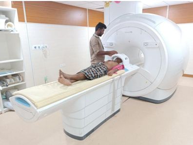
1.5 TESLA MRI
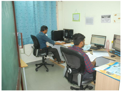
MRI REPORTING ROOM
8) Interventional Procedures:
We have a dedicated suite for performing interventrion procedures in our department.
More than 3 to4 procedures are routinely done per day.
Biopsy guns with co axial needles are available.
Ultrasound guided biopsies , ultrasound guided aspirations of cysts, abscess of solid organs and body cavities are routinely done .
Ultrasound guided pigtail insertion for liver abscess, pyonephrosis are routinely done.
Ultrasound guided thyroid and breast FNAC
CT guided biopsies and pigtail insertion are routinely done.
All the X ray units and CT machines in our department has obtained appropriate license from the atomic energy regulatory board and isappropriately functioning.
All the ultrasound machines in our department are being registered under the PCPNDT ACT.
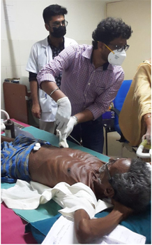
USG Guided Intervention
Clinical Workload In The Past 3 Years:
| Particulars | Year 1 2019 |
Year 2 2020 |
Year 3 2021 |
|||
|---|---|---|---|---|---|---|
| Conventional Radiology (X-Rays.) | 125087 | 68227 | 152611 | |||
| Contrast Radiology (Barium / IVP) | 1537 | 462 | 1192 | |||
| USGs (Gray Scale) | 64352 | 28613 | 36256 | |||
| USGs (Colour Doppler) | 2635 | 2592 | 1983 | |||
| USG guided FNAC / Aspirations etc. | 127 | 20 | 424 | |||
| CT Scans | 49258 | 53252 | 79875 | |||
| CT guided FNAC / Biopsies | 25 | 4 | 93 | |||
| MRI (Plain / contrast) | 6608 | 280 | 3913 | 203 | 5422 | 347 |
| Angiography (conventional/DSA) | 24 | 9 | 10 | |||
| Mammography | 229 | 192 | 141 | |||
| Any other investigation | - | - | - | |||
Daily Average Census Of Various Sections Of Our Department:
| 1. | X-Rays | 411 |
| 2. | Special procedures (IVP etc.) | 6 |
| 3. | Ultrasonography and Doppler | 183 |
| 4. | C.T. Scans | 230 |
| 5. | MRI scans | 25 |
| 6. | Mammography | 4 |
| 7. | Intervention | 4 |
Special Clinics Conducted By Our Department:
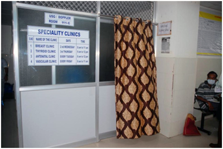
| Name of the Clinic | Weekday/s | Timings | Name of Clinic In-charge |
|---|---|---|---|
| Vascular clinic | Every Friday | 8AM-11AM | Dr.Elanchezhian |
| Antenatal clinic | Every Tuesday | 8AM-12PM | Dr.Dheepa |
| Breast clinic | 2nd Wednesday | 8AM-11AM | Dr.Jaysiya |
| Thyroid clinic | 3rd Thursday | 9AM-12PM | Dr.Ramachandran |
Radiation Protective Measures
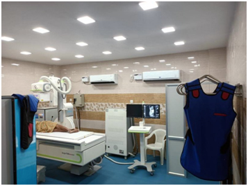
Full time RSO (Radiation supervising Officer) cum Physicist is posted in Radio-diagnosis department. He is taking care of Machine, Staff and Patient Radiation safety.
All the CT and XRAY units are installed and certified under AERB guidelines.
Thermo luminscent dosimeter - TLD badges are available to all staff.
Protective measures is available as per BARC specification
Lead protective screens, Lead apron, Thyroid and Gonadal Guards, Lead goggles, gloves are available.
Department Library And Museum:
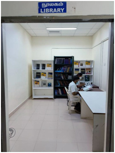
A well furnished department library with internet facilities , latest books an djournals are available.
| Total No. of Books | 197 |
|---|---|
| Purchase of latest editions in past 3 years | 94 |
| Number of Journals | 18 |
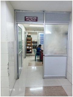
A well spacious museum with more than 300 specimens and more than 20 charts and diagrams are available.
Academic Activities:
First CRA course was started in 2008.
First DRA course was started in 2008
First DRDT Course started in 2011
Ultrasound training is given to the PHC medical officers, Government hospitalAsst.surgeonsand ESI hospital doctors throughout the calendar year.
Basic Radio-diagnosis are classes taken for the Pre-final, final year M.B; B.S students andPost graduate students (Gen.Sur., Gen.Med.,Ortho, OG) during their Radio-diagnosis posting.
PCPNDT six month USG training given to service and non-service medical officer fromDec2015 to May 2016
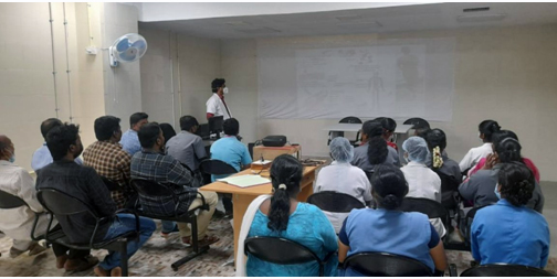
Theory classes are being taken to prefinal MBBS students every Tuesday 4-5pm second half of every year from july to december.
Post graduate MD Radiodiagnosis degree course with an intake of 3 seats per year is sanctioned from the academic year 2022
Post Graduate (PG ) course:
MD Radiodiagnosis degree course with 2 seats is started in the academic year 2022 - 2023
Periodic academic activities are done in our department. Morning classes are being taken routinely by our assistant professors from 8am to 9am.
| S.no | Academic activities | Schedule |
|---|---|---|
| 1 | Theory class | Daily morning 8am to 9am |
| 2 | Clinical seminars | Every Wednesday |
| 3 | Journal clubs | Every saturday |
| 4 | Tutorials | Afternoon 12pm to 1pm |
| 5 | Film reading session | Daily afternoon 12pm to 1pm |
| 6 | Guest lectures | Yearly thrice |
Under Graduate Course:
15 days OP posting during 2nd clinical year and theory classes as per revised teaching schedule are taken to the prefinal year students.
UG (MBBS Pre Final Year) Theory Class Schedule
| S.no | Topic |
|---|---|
| 1 | X-RAY BASIC AND SAFETY MEASURES |
| 2 | FLUROSCOPY/IITV BASIC |
| 3 | CHEST X-RAY |
| 4 | ABDOMINAL X-RAY |
| 5 | X-RAY PROCEDURES BARIUM SWALLOW,MEAL,ENEMA,LOOPOGRAM |
| 6 | X-RAY PROCEDURES IVU,AUG,MCU,PCN,FISTULOGRAM,SINOGRAM,SIALOGRAM |
| 7 | USG BASIC,PATIENT PREPARATION,INDICATION |
| 8 | FOCUS USG IN EMERGENCY EMERGENCY USES OF USG |
| 9 | OBS ULTRASOUND BASICS |
| 10 | DOPPLER BASICS |
| 11 | CT BASICS |
| 12 | CT BRAIN ANATOMY AND PATHOLOGY |
| 13 | CT ABDOMEN- PATIENT PREPARATION,SCAN,ANATOMY |
| 14 | CT-MPR AND ANGIOGRAM |
| 15 | MRI BASICS,SEQUENCES,SAFETY MEASURES |
| 16 | MRI BRAIN |
| 17 | MRI BASIC ABDOMEN,PELVIS |
| 18 | USG –SMALL PARTS BASIC |
| 19 | CONTRAST MEDIA |
| 20 | NUCLEAR MEDICINE RADIO ACTIVITIES,TAGGING MATERIALS,SAFETY MEASURES |
| 21 | VENTILATION PERFUSION SCAN BONE SCAN |
| 22 | RADIATION DOSE,RISK AND SAFETY |
| 23 | ANATOMICAL AND FUNCTIONAL IMAGING, PET,FUSION IMAGING |
| 24 | CONTRAST MEDIA REACTIONS |
Paramedical Course :
DRDT course - average intake of 10students per year.
The course duration is 2years
The course is affiliated to Tamil nadu Dr. MGR medical university.
The academic curriculum includes providing
- Basic training in operations of X-ray, CT, MRI, Ultrasound and understanding the basic physics.
- Contrast investigations -Setting protocols for methods of selecting cases, performing the procedure, Statistical correlation, Maintenance of Records, Collection of cases for Case Records and Post procedural care
- Basics of Anesthesia, Sterility & drugs used in Radiology Department, Barium, Ionic& Non-ionic contrast medium, Gadolinium, Atropine, Local anesthetics IV analgesics ,vasodilator, Thrombolytic drugs, Drugs used in Resuscitation, H1 and H2 blockers
-MRI -Basics of MRI, Equipment handling
Student assessment is done by conducting theory examination every fortnight.
Research Papres And Publications:
| S.no | Faculty Name | Details of publication: |
|---|---|---|
| 1 | DR.P.KUMAR MDRD., | Kumar P ,Arun C . MRI colonography versus conventional colonscopy in detection of colonic polyposis. International journal of latest research in science and technology, march – april 2016 ; volume(5) Issue(2) :page number 80-85.Available from: http://www.mnkjournals.com/ijlrst.htm |
| ArunC ,Kumar P .Utility of Transrectal ultrasound with power Doppler imaging guided biopsy in the detection of prostate cancer. International journal of latest research in science and technology, march – april2016 ; volume(5) Issue(2) :page number 40-43. Available from: https://www.mnkpublication.com/journal/ijlrst/index.php | ||
| Kumar P ,Arun C . MRI Evaluation of ligamentous injuries and meniscal injuries of the knee joint . International journal of science and research, February 2019 ; volume(8) Issue(2) :page number 2245-2249 .Available from : www.ijsr.net | ||
| Kumar P ,Arun C . Evaluation of Fistula in Ano MR Fistulography. International journal of science and research, February 2019 ; volume(8) Issue(2) :page number 2250-2254 .Available from : www.ijsr.net | ||
| 2 | DR.N.SUNDARESWARAN MDRD., | SundareswaranN ,Babu peter , Kailasanathan .Inphase opposed phase sequence in MRI differentiating neoplastic lesions of bone marrow from non neoplastic lesions of bone marrow . International journal of latest research in science and technology, January-February 2016 ; volume(5) Issue(1) :page number 146-156 . Available from: http://www.mnkjournals.com/ijlrst.htm |
| ShankaranandhR ,Sundareswaran N .Characterization of Adrenal masses with contrast enhanced CT-washout study . International journal of latest research in science and technology, May-June 2016 ; volume(5) Issue(3) :page number 6-10 .Available from: http://www.mnkjournals.com/ijlrst.htm | ||
| KalaivaniP ,Sundareswaran N .Hippocampal abnormalities in Adults with unilateral temporal lobe Epilepsy- A Diffsuion Tensor imaging study. International journal of science and research, November 2019 ; volume(8) Issue(11) :page number 1564-1571 .Available from : www.ijsr.net | ||
| SundareswaranN ,Elanchezhian E .Role of time intensity curve in dynamic contrast MRI evaluation of soft tissue tumour. International journal of contemporary medicine surgery and Radiology,April-June 2020 ;volume(5) Issue(2): page number B60-B65 .Available from: http://dx.doi.org/10.21276/ijcmsr.2020.5.2.14 | ||
| 3 | DR.E.ELANCHEZHIAN MDRD., | KalaivaniP ,Elanchezhian E . Extratemporal abnormalities in adults with unilateral temporal lobe epilepsy –A diffusion tensor imaging study. International journal of Radiology and Diagnostic Imaging, March - 2020 ; volume(3) Issue(2): page number 9-17. Available from: http://dx.doi.org/10.33545/26644436.2020.v3.i2a.90 |
| ElanchezhianE ,Kalaivani P .MRI findings in children with global developmental delay. International journal of Radiology and Diagnostic Imaging, March - 2020 ; volume(3) Issue(2): page number 7-8. Available from: http://dx.doi.org/10.33545/26644436.2020.v3.i2a.89 | ||
| SundareswaranN ,Elanchezhian E .Role of time intensity curve in dynamic contrast MRI evaluation of soft tissue tumour. International journal of contemporary medicine surgery and Radiology,April-June 2020 ;volume(5) Issue(2): page number B60-B65 . Available from: http://dx.doi.org/10.21276/ijcmsr.2020.5.2.14 | ||
| 4 | DR.M.SANKAR MDRD., | SankarM ,Vidhya E .Descriptive study on various perianal fistulas in rural population using MR Fistulogram. International journal of Radiology and Diagnostic Imaging, February-2021;volume(4) Issue(1): page number 228-230. Available from: http://dx.doi.org/10.33545/26644436.2021.v4.i1d.189 |
| VidhyaE,SankarM.Correlation of CT portography vasculature to child pugh classification of cirrhotic patients. International journal of Radiology and Diagnostic Imaging, February-2021;volume(4) Issue(1): page number 231-233. Available from: http://dx.doi.org/10.33545/26644436.2021.v4.i1d.190 | ||
| 5 | DR.E.VIDHYA DMRD., DNB., | SankarM ,Vidhya E .Descriptive study on various perianal fistulas in rural population using MR Fistulogram. International journal of Radiology and Diagnostic Imaging, February-2021 ;volume(4) Issue(1): page number 228-230. Available from: http://dx.doi.org/10.33545/26644436.2021.v4.i1d.189 |
| VidhyaE,SankarM.Correlation of CT portography vasculature to child pugh classification of cirrhotic patients. International journal of Radiology and Diagnostic Imaging, February-2021;volume(4) Issue(1): page number 231-233. Available from: http://dx.doi.org/10.33545/26644436.2021.v4.i1d.190 |
Department Census Details:
| Department Of Radiology Census | ||||||||||||||||||||||||||
|---|---|---|---|---|---|---|---|---|---|---|---|---|---|---|---|---|---|---|---|---|---|---|---|---|---|---|
| YEAR | X RAY | MRI | CT angio | CT | USG | Doppler | USG guiderd Intervention | CT / guiderd Intervention | ||||||||||||||||||
| CASES | Total exposure | Total films | Contrast xray | |||||||||||||||||||||||
| OP | IP | Mamo | Total | BAR | IOD | Total | OP | IP | Con | Total | OP | IP | Con | Total | OP | IP | AN case | Duty USG | Total | |||||||
| 2012 | 2455 | 31575 | 65 | 34095 | 74879 | 8956 | 430 | 818 | 1248 | 1122 | 1692 | -- | 2814 | 117 | 6400 | 9834 | 1764 | 17998 | 8259 | 12414 | 3276 | 11851 | 35800 | 1520 | 122 | 28 |
| 2013 | 24813 | 31575 | 83 | 56471 | 74156 | 11525 | 386 | 932 | 1318 | 2374 | 1491 | 197 | 4062 | 111 | 7620 | 12200 | 2239 | 22059 | 13271 | 16962 | 3380 | 8565 | 42178 | 1476 | 116 | 24 |
| 2014 | 28822 | 32336 | 87 | 61245 | 77873 | 16852 | 428 | 907 | 1335 | 2793 | 1787 | 240 | 4820 | 95 | 9413 | 16565 | 4251 | 30229 | 16735 | 13556 | 3230 | 10252 | 43773 | 1986 | 130 | 16 |
| 2015 | 26497 | 36980 | 40 | 63517 | 45369 | 12654 | 472 | 858 | 1330 | 2588 | 1882 | 400 | 4870 | 89 | 8474 | 16855 | 3404 | 28733 | 14293 | 15642 | 3492 | 11125 | 44552 | 1640 | 136 | 20 |
| 2016 | 46190 | 54385 | 3 | 103925 | 221316 | 43414 | 561 | 963 | 1524 | 2803 | 2452 | 289 | 5544 | 47 | 8251 | 22394 | 7092 | 37737 | 15263 | 19398 | 1218 | 13825 | 49704 | 1110 | 122 | 21 |
| 2017 | 42088 | 46240 | 97 | 88425 | 114080 | 11763 | 539 | 893 | 1432 | 2978 | 2564 | 164 | 5706 | 105 | 9347 | 25203 | 2914 | 37464 | 14970 | 28788 | 3367 | 17115 | 64240 | 1765 | 110 | 21 |
| 2018 | 47890 | 55785 | 250 | 103925 | 221316 | 13071 | 325 | 803 | 1128 | 3996 | 2553 | 249 | 6798 | 118 | 14336 | 27249 | 3001 | 44586 | 19529 | 19454 | 6157 | 13825 | 58965 | 3061 | 122 | 20 |
| 2019 | 55170 | 69688 | 229 | 125087 | 276773 | 17046 | 528 | 1009 | 1537 | 3772 | 2846 | 280 | 6898 | 132 | 24537 | 21803 | 2862 | 49202 | 16710 | 22415 | 7103 | 18129 | 64357 | 2635 | 131 | 25 |
| 2020 | 20201 | 48026 | 192 | 68419 | 140570 | 12470 | 149 | 313 | 462 | 1495 | 2418 | 203 | 4116 | 115 | 29187 | 22217 | 1848 | 53252 | 4050 | 10665 | 5339 | 8559 | 28613 | 2592 | 20 | 4 |
| 2021 | 19690 | 46691 | 141 | 66522 | 152613 | 15158 | 426 | 766 | 1192 | 2922 | 2500 | 347 | 5769 | 190 | 46644 | 30441 | 2790 | 79875 | 7653 | 17030 | 5844 | 5729 | 36256 | 1983 | 424 | 93 |
CMHIS Beneficiaries Detail:
| YEAR | CT | MRI | 128SLICE CT ANGIO | TOTAL AMOUNT GENERATED |
|---|---|---|---|---|
| 2012 | 10 | 16 | 01 | 46050 |
| 2013 | 11 | 46 | 02 | 124400 |
| 2014 | 760 | 447 | 03 | 1408500 |
| 2015 | 1797 | 2329 | 09 | 6328000 |
| 2016 | 1353 | 2465 | - | 6837500 |
| 2017 | 905 | 2651 | 30 | 7097900 |
| 2018 | 1061 | 3357 | 49 | 8997800 |
| 2019 | 958 | 2969 | 43 | 9467500 |
| 2020 | 349 | 990 | 22 | 2896500 |
| 2021 | 275 | 2081 | 53 | 5640000 |
| 2022 | 904 | 2611 | 175 | 80,95,000 |
COVID CT Census For The Year 2021:
| GOVERNMENT MOHAN KUMARAMANGALAM MEDICAL COLLEGE SALEM DEPARTMENT OF RADIOLOGY | |
|---|---|
| COVID CT CENSUS FOR THE YEAR OF 2021 | |
| MONTH | NO OF CT CASES |
| JANUARY | 812 |
| FEBRUARY | 582 |
| MARCH | 620 |
| APRIL | 3569 |
| MAY(UPTO 28/05/2021) | 6015 |
| TOTAL | 11598 |
CME/WORKSHOPS/CONFERENCES:
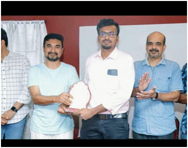
Assistant professor DR. Gomathiponshankar MDRD., receiving
momento for his lecture in salem subchapter IRIA meet - 2022
held at yercaud.
Image Galleries:
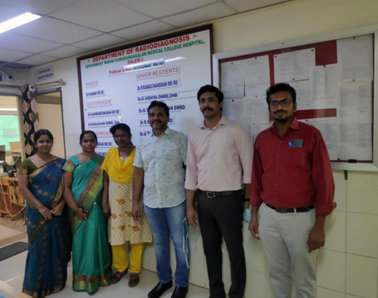
PHC medical officers on completion of 6months
antenatal ultrasound training
